Questões de Concurso
Sobre radiologia em odontologia
Foram encontradas 2.116 questões


Dentre suas muitas vantagens, encontra-se:
Sobre a interpretação radiográfica da face, analise as afirmativas e assinale V para as verdadeiras e F para as falsas.
( ) O maior número de estruturas anatômicas aparece nas radiografias da mandíbula.
( ) O conduto incisivo, através do qual passam os nervos nasopalatinos e o ramo anterior dos vasos palatinos descendentes, nem sempre é observado na radiografia.
( ) A distância entre as fossas nasais e as raízes dos dentes anteriores não varia muito nas radiografias de paciente para paciente.
( ) A hipercementose é observada na radiografia como um alargamento bulboso contornado por um espaço contínuo do ligamento periodontal e uma lâmina dura normal sem interrupções.
Assinale a sequência CORRETA.
“A técnica radiográfica _______________________ permite visualizar a base do crânio, avaliar o posicionamento das cabeças da mandíbula, inves\gar o seio esfenoidal e a parede lateral dos seios maxilares. Sua maior importância, no entanto, está na detecção de eventuais fraturas do arco zigomático.”
(Boracks, 2011.)
Assinale a alternativa que completa corretamente a afirmativa anterior.
A maioria dos esquemas de classificação dos dentes impactados baseia-se na radiografia panorâmica, visando prever problemas pré e pós-operatórios. Sobre o sistema de Pell e Gregory para classificar terceiros molares impactados, marque V para as afirmativas verdadeiras e F para as falsas.
( ) Em relação à margem anterior do ramo, os dentes que se apresentam posicionados posteriormente de maneira que, aproximadamente, metade esteja coberto pelo ramo, a relação do dente com o ramo será de classe 3.
( ) Em relação à margem anterior do ramo, os dentes que apresentam diâmetro mesiodistal da coroa completamente anterior à margem anterior do ramo da mandíbula classifica-se como relação classe 1.
( ) Em relação ao plano oclusal, uma impacção classe C é aquela na qual a superfície oclusal do dente impactado está abaixo da linha cervical do segundo molar.
( ) Em relação ao plano oclusal, uma impacção classe B é aquela na qual a superfície oclusal do dente impactado está à altura ou próxima ao nível do plano oclusal do segundo molar.
A sequência está correta em
No planejamento cirúrgico dos dentes não irrompidos, o exame radiográfico é de capital importância, sobretudo no tocante à localização dos mesmos. Em relação aos métodos radiográficos para auxiliar a localização dos dentes não irrompidos, analise as afirmativas.
I. O método de Miller-Winter emprega o princípio da tripla incidência. Originalmente, este método foi idealizado para localizar os dentes não irrompidos da região dos molares inferiores.
II. O método de localização radiográfica de Donovan emprega o princípio da dupla incidência. Este método de localização radiográfica visa auxiliar a localização de dentes localizados na região mais posterior de mandíbula, possibilitando a visualização da região radicular do dente não irrompido.
III. O método de localização radiográfica de Parma emprega o princípio da dupla incidência para encontrar dentes não irrompidos na região de molares inferiores. O método emprega uma radiografia intrabucal periapical da região dos dentes molares inferiores e uma radiografia oclusal direta com filme periapical.
IV. Ao utilizar a técnica de Clark, onde se realizou um desvio distorradial, observou-se que a imagem radiográfica de um canino superior não irrompido apresenta a mesma posição em relação à tomada radiográfica periapical da região. Tal situação indica que o canino superior está situado equidistante do vestíbulo e do palato.
Estão corretas apenas as afirmativas
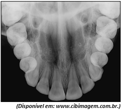
Qual é o tipo de radiografia apresentada?

 Conheça nossos planos